Confocal and Light Sheet imaged Embryonic cells#
Ascadian Embryo#
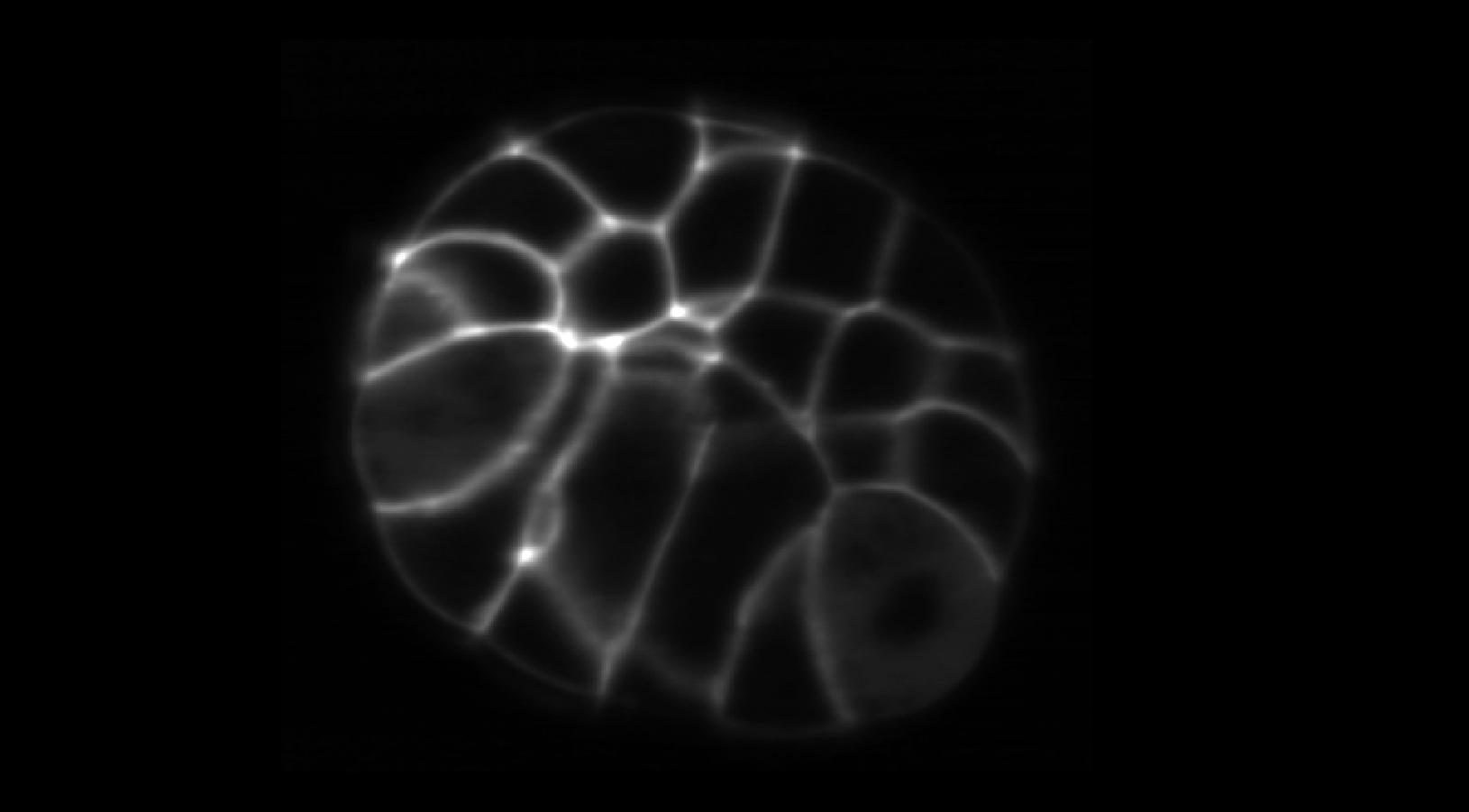
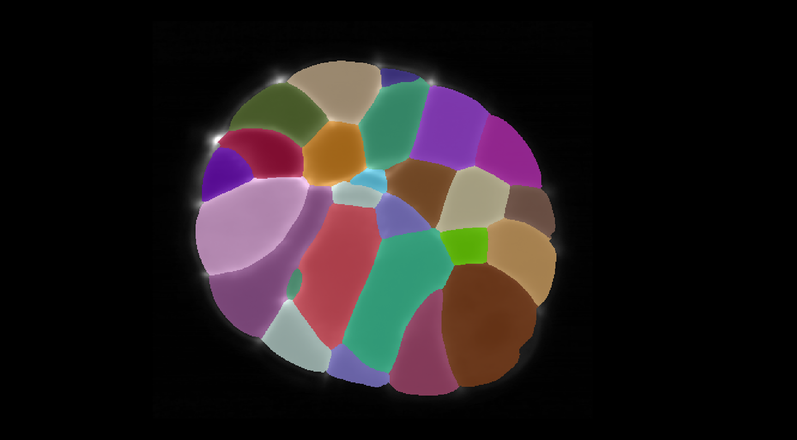
In this example we consider a dataset imaged using Light sheet fused from four angles to create a single channel 3D image of Phallusia Mammillata Embryo created using live SPIM imaging. The training data can be found here. For this imaging modality we trained only a UNet model to segment the interior region of the cells and by using slice_merge=True and
expand_labels=True in the VollSeg parameter setting we obtained the following segmentation result along with the metrics compared to the ground truth.
Raw Ascadian Embryo |
Prediction Ascadian Embryo |
|---|---|
|
|


